
Missed Root Canal Symptoms in 2026
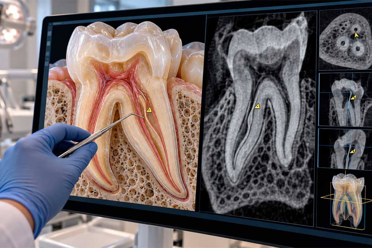
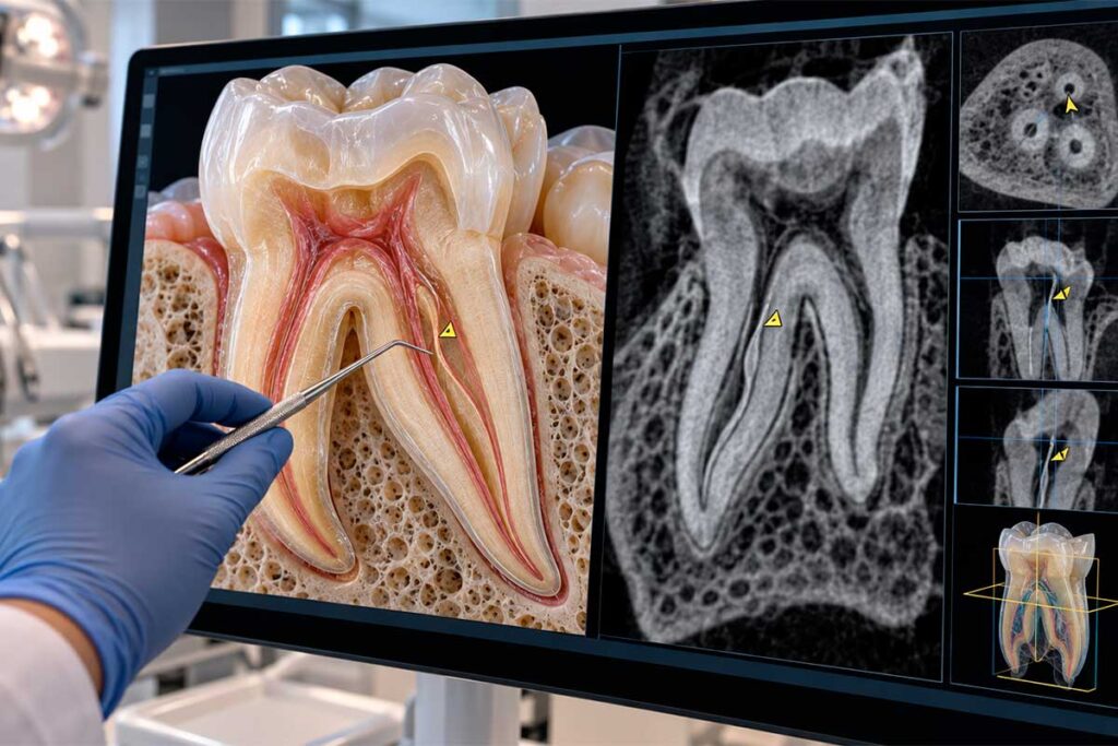
A root canal system is rarely as simple as a single straight tube inside a tooth. Roots can contain narrow branches, curved pathways, connections between canals, calcified areas, and additional canals that are difficult to identify on conventional two-dimensional dental images. That anatomical complexity helps explain why a previously treated tooth sometimes requires a more detailed evaluation.
Patients searching for missed root canal symptoms are often experiencing continued discomfort after treatment or symptoms that returned after a period of improvement. An untreated canal is one possible explanation, but symptoms alone cannot establish that diagnosis. Cracks, reinfection, bite-related forces, periodontal problems, structural damage, or inflammation around the root can create similar complaints.
At Nuvo Dental in Orlando, Dr. Pedro Ramirez approaches these situations as diagnostic problems rather than assuming that a previous procedure has failed. The objective is to understand the treatment history, current symptoms, tooth structure, imaging findings, and internal anatomy before deciding whether observation, additional testing, restorative treatment, retreatment, or another form of care deserves consideration.
Can a Tooth Have More Root Canals Than Expected?
Yes. Teeth have recognizable anatomical patterns, but those patterns include considerable natural variation. Two upper first molars can look almost identical from the outside while their internal root canal systems differ substantially.
One clinically important example is the second mesiobuccal canal, commonly called MB2, in an upper molar. Research has repeatedly shown that this canal is common rather than exceptional. A 2026 systematic review and meta-analysis of South American CBCT studies reported an overall MB2 prevalence of 59%, reaching 66% in maxillary first molars.
Earlier global CBCT research also demonstrated wide geographic variation and found MB2 anatomy in a large proportion of maxillary first molars. These findings reinforce a practical lesson: clinicians need to anticipate anatomical variation rather than expect every tooth to conform to a simplified diagram.

Why Root Canal Anatomy Can Be Difficult to Identify
The challenge is not simply counting canals. A canal can be extremely narrow, partially calcified, sharply curved, or positioned in a way that makes its entrance difficult to visualize. Some root canal systems contain branches and communications that become apparent only through careful exploration and appropriate imaging.
Conventional dental radiographs remain valuable because they provide information about roots, previous treatment, bone changes, and surrounding structures. Their limitation is dimensional: a three-dimensional tooth is being represented in two dimensions, so anatomical structures can overlap.
This is one reason endodontic diagnosis depends on combining several sources of information. Symptoms, clinical tests, previous radiographs, current imaging, access anatomy, restorative condition, and the clinician’s interpretation all contribute different pieces of the case.
Accessory Canals and Anatomical Variations
Accessory anatomy can include additional main canals, lateral branches, fins, isthmuses, and other internal connections. These structures vary in size and clinical significance, and they are not equally visible with every diagnostic method.
The existence of anatomical variation does not mean every small branch must be identified as an independent canal during routine treatment. The clinically relevant question is whether untreated anatomy could be harboring tissue or microorganisms that help explain persistent disease.
What Is an MB2 Canal?
“MB2” refers to a second canal associated with the mesiobuccal root of an upper molar. The mesiobuccal root frequently contains one canal, but a second pathway may be present and can vary greatly in visibility, curvature, size, and configuration.
The clinical relevance comes from its frequency and the difficulty it can sometimes present. A systematic review of CBCT-based prevalence studies found MB2 in about 69.6% of maxillary first molars and 39% of second molars across the populations included.

What Are the Possible Missed Root Canal Symptoms?
People often search for a definitive set of symptoms that proves an extra canal was left untreated. Dentistry cannot provide that level of certainty from symptoms alone because several conditions can create overlapping patterns of pain or inflammation.
Findings that may justify endodontic re-evaluation include:
Persistent or recurrent discomfort in a previously treated tooth, particularly when symptoms continue beyond the expected post-treatment period.
Tenderness or pain when biting or releasing pressure, especially if the symptom persists or becomes more noticeable.
Swelling, drainage, a gum pimple near the treated tooth, or other signs that may indicate ongoing infection.
Symptoms that disappear after treatment and return weeks, months, or years later.
A new radiographic finding around a root or a previously treated tooth that requires further interpretation.
An unexplained change in the way the tooth feels despite apparently completed root canal treatment.
Why Persistent Pain Does Not Automatically Mean a Canal Was Missed
Root canal treatment addresses disease inside the pulp and root canal system, but the treated tooth remains connected to periodontal tissues, bone, the bite, and surrounding structures. Pain originating in those areas can resemble endodontic pain.
A tooth can also have more than one problem simultaneously. A patient may have received technically adequate root canal treatment and later develop a fracture, recurrent decay, leakage around a restoration, or excessive biting pressure.
Temporary post-treatment tenderness is another consideration. Mild sensitivity during chewing can occur as the tissues around the root recover. Persistent, worsening, severe, or recurrent symptoms require a different level of attention because the clinician needs to determine whether healing is progressing normally or another problem is present.

When a Previously Treated Tooth May Need Re-Evaluation
Re-evaluation is appropriate when symptoms persist beyond what would normally be expected, return after a period of stability, or are accompanied by new clinical or radiographic findings. It is also reasonable when a tooth has complex anatomy and the existing records do not fully explain the condition.
Re-evaluation does not automatically lead to retreatment. Depending on the findings, the next step may involve monitoring, adjusting the bite, treating a restorative issue, obtaining additional imaging, considering endodontic retreatment, evaluating a fracture, or referring the patient for additional specialty care.
That diagnostic sequence protects the patient from assuming that every post-treatment symptom means the previous root canal must be repeated.
Choosing Endodontic Care in Orlando
Persistent symptoms after root canal treatment can be frustrating because patients often expect the issue to be completely resolved once the procedure is finished. When discomfort remains or returns, the first priority should be identifying the cause rather than assuming treatment failure.
Nuvo Dental’s Endodontics care can help evaluate the biological, anatomical, structural, and restorative factors surrounding a symptomatic tooth. Complex internal anatomy is one possibility, but the final treatment plan should reflect the complete clinical picture.
For patients in Orlando who are concerned about pain after previous root canal therapy, a focused evaluation with Dr. Pedro Ramirez can help determine whether additional anatomy may be involved, whether another condition better explains the symptoms, and what next step is appropriate for the tooth.
FAQS
Why does my tooth still hurt after a root canal?
Several explanations are possible. Temporary tenderness can occur during healing, while persistent or recurrent pain may be related to ongoing endodontic disease, a crack, reinfection, bite-related pressure, periodontal issues, structural damage, or another source of pain. An examination is necessary to distinguish among them.
Can a tooth really have an extra root canal?
Yes. Root canal anatomy varies naturally. Upper molars frequently have an additional MB2 canal, while lower molars may occasionally contain extra mesial anatomy. The number and configuration of canals can differ between patients and even between corresponding teeth in the same mouth.
Can a root canal fail because an extra canal was missed?
Yes, untreated canal anatomy can contribute to persistent or recurrent endodontic disease because tissue or microorganisms may remain within areas that were not adequately treated. It is one possible explanation among several, so diagnosis should come before retreatment.
What are the most common missed root canal symptoms?
Possible missed root canal symptoms can include persistent pain, tenderness when biting, swelling, drainage, or symptoms that return after a period of improvement. These findings are not specific to a missed canal and require professional evaluation to determine the cause.
